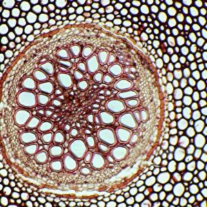
Fern rhizome, light micrograph

![]()
Wall Art and Photo Gifts from Science Photo Library
Fern rhizome, light micrograph
Fern rhizome, light micrograph. Transverse section through the central portion of the underground stem (rhizome) of the bracken fern (Pteridium aquilinum). The outer epidermis (black) covers a thin cortex layer (white) with parenchyma cells (green) below that. In the centre of the stem are vascular strands (oval-round) called meristeles. These include wood cells (xylem, red). The cells (black) between the meristeles are fibrous supporting tissue. Magnification: x11 when printed at 10 centimetres wide
Science Photo Library features Science and Medical images including photos and illustrations
Media ID 6308731
© DR KEITH WHEELER/SCIENCE PHOTO LIBRARY
Cellular Cortex Cross Section Epidermis Fern Ferns Internal Structure Parenchyma Part Parts Plant Anatomy Pteridium Aquilinum Pteridophyte Pteridophytes Rhizome Structural Tissue Transverse Vascular Xylem Cells Light Micrograph Light Microscope Section Sectioned
MADE IN THE USA
Safe Shipping with 30 Day Money Back Guarantee
FREE PERSONALISATION*
We are proud to offer a range of customisation features including Personalised Captions, Color Filters and Picture Zoom Tools
SECURE PAYMENTS
We happily accept a wide range of payment options so you can pay for the things you need in the way that is most convenient for you
* Options may vary by product and licensing agreement. Zoomed Pictures can be adjusted in the Cart.