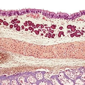
Nose mucosa, light micrograph

![]()
Wall Art and Photo Gifts from Science Photo Library
Nose mucosa, light micrograph
Nose mucosa. Light micrograph of a section through the nasal mucosa (the tissue that lines the airways of the nose), in the region lying over the nasal concha. The nasal concha is a bony region that is responsible for forcing inhaled air to flow through hair-like cilia. The cilia form the upper surface of the columnar epithelium cells (orange, top) that make up the top layer of the nasal mucosa. The cilia are coated in sticky mucus produced by goblet cells in the membrane, which serves to trap dust and pathogens. Under the epithelium is the glandular and vascular lamina propria (green and purple). Magnification: x300 when printed 10cm wide
Science Photo Library features Science and Medical images including photos and illustrations
Media ID 6449361
© STEVE GSCHMEISSNER/SCIENCE PHOTO LIBRARY
Ciliated Epithelial Epithelium False Colour Glands Goblet Histological Histology Lamina Propria Mucosa Mucous Mucous Membrane Mucus Nasal Nasal Cavity Nose Olfaction Olfactory Sense Smell Cells False Coloured Light Micrograph Light Microscope
EDITORS COMMENTS
This print showcases the intricate beauty of the nose mucosa, providing a fascinating glimpse into the inner workings of our respiratory system. The light micrograph captures a section through the nasal mucosa, revealing the tissue that lines the airways of our nose. Specifically, it focuses on the region above the nasal concha – a bony structure responsible for directing inhaled air through hair-like cilia. The columnar epithelium cells form an orange upper layer and are adorned with delicate cilia that play a crucial role in filtering incoming air. These cilia are coated with sticky mucus produced by goblet cells within the membrane. This ingenious mechanism serves to trap dust particles and pathogens, safeguarding our respiratory health. Beneath this protective layer lies the glandular and vascular lamina propria depicted in vibrant shades of green and purple. This underlying layer supports various functions necessary for maintaining optimal nasal function. With its false-colored presentation highlighting key cellular components, this image not only offers aesthetic appeal but also serves as an invaluable tool for studying histology and understanding how our sense of smell operates at a microscopic level. Displayed at x300 magnification when printed 10cm wide, this remarkable photograph from Science Photo Library provides us with an awe-inspiring glimpse into one small aspect of human biology – reminding us once again of nature's complexity and beauty hidden within even seemingly ordinary parts of our bodies.
MADE IN THE USA
Safe Shipping with 30 Day Money Back Guarantee
FREE PERSONALISATION*
We are proud to offer a range of customisation features including Personalised Captions, Color Filters and Picture Zoom Tools
SECURE PAYMENTS
We happily accept a wide range of payment options so you can pay for the things you need in the way that is most convenient for you
* Options may vary by product and licensing agreement. Zoomed Pictures can be adjusted in the Cart.