Trachea lining, TEM C014 / 1471

![]()
Wall Art and Photo Gifts from Science Photo Library
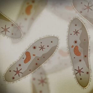
Trachea lining, TEM C014 / 1471
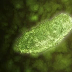
Trachea lining. Transmission electron micrograph (TEM) of a transverse section through the lining of the trachea (windpipe), which links the larynx (voicebox) to the lungs. The epithelial lining consists of cells that have hair-like cilia (red, upper left) on their surface and mucus-secreting glands. Mucus secreted by the glands traps debris, such as dust particles or bacteria, in the inhaled air, while the beating of the cilia moves the mucus and particles upwards out of the respiratory tract, helping to keep the lungs and airways clear and prevent infection. Clear basal bodies (red rings) and mitochondria (turquoise) are visible in the cell bodies. Magnification: x7000 when printed 10 centimetres wide
Science Photo Library features Science and Medical images including photos and illustrations
Media ID 9227079
© STEVE GSCHMEISSNER/SCIENCE PHOTO LIBRARY
Bodies Cilia Cilium Cytological Cytology Gland Glands Histological Histology Lining Microvilli Microvillus Mitochondria Mitochondrion Mucous Cell Mucus Respiratory System Secretory Trachea Transmission Electron Micrograph Transmission Electron Microscope Transverse Section Windpipe Sectioned
EDITORS COMMENTS
This print showcases the intricate lining of the trachea, captured through a transmission electron microscope. The trachea, also known as the windpipe, is responsible for connecting the larynx to the lungs and plays a vital role in our respiratory system. In this transverse section image, we observe the epithelial lining composed of specialized cells adorned with hair-like cilia on their surface. These red-colored cilia are crucial in maintaining healthy lungs and airways by constantly beating and moving mucus along with trapped debris upwards out of our respiratory tract. The mucus-secreting glands depicted here play an essential role in trapping harmful particles like dust or bacteria present inhaled air. This protective mechanism prevents infections and ensures that our lungs remain clear from any potential threats. Upon closer inspection, clear basal bodies resembling red rings can be observed within cell bodies alongside turquoise mitochondria. These structures contribute to cellular functions such as energy production and organization within these specialized cells. With a magnification of x7000 when printed at 10 centimeters wide, this remarkable photograph offers us a glimpse into the microscopic world of biology and anatomy. It serves as a reminder of how intricately designed our human body is, highlighting its fascinating mechanisms for self-preservation and protection against external elements.
MADE IN THE USA
Safe Shipping with 30 Day Money Back Guarantee
FREE PERSONALISATION*
We are proud to offer a range of customisation features including Personalised Captions, Color Filters and Picture Zoom Tools
SECURE PAYMENTS
We happily accept a wide range of payment options so you can pay for the things you need in the way that is most convenient for you
* Options may vary by product and licensing agreement. Zoomed Pictures can be adjusted in the Cart.