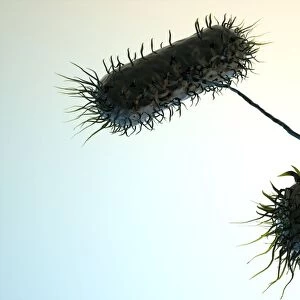
Gall bladder surface, SEM

![]()
Wall Art and Photo Gifts from Science Photo Library
Gall bladder surface, SEM
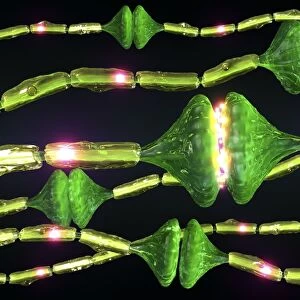
Gall bladder. Coloured scanning electron micrograph (SEM) of the internal surface of a gall bladder. This mucosa lining is made up of columnar epithelial cells (green and yellow). Each cell of the gall bladder lining has microvilli (tiny projections) that increase its surface area and aid water uptake. Connective tissue (brown) is seen below the lining where some of the cells have come away. The gall bladder is a sac that concentrates and stores bile, produced by the liver, and releases it into the duodenum (small intestine), where it aids the digestion of fats. Magnification: x55 when printed 10 centimetres wide
Science Photo Library features Science and Medical images including photos and illustrations
Media ID 6422684
© STEVE GSCHMEISSNER/SCIENCE PHOTO LIBRARY
Bile Columnar Connective Tissue Digestion Digestive Epithelial False Colour Gall Bladder Histological Histology Microvilli Microvillus Mucosa Storage Surface Cells False Coloured
MADE IN THE USA
Safe Shipping with 30 Day Money Back Guarantee
FREE PERSONALISATION*
We are proud to offer a range of customisation features including Personalised Captions, Color Filters and Picture Zoom Tools
SECURE PAYMENTS
We happily accept a wide range of payment options so you can pay for the things you need in the way that is most convenient for you
* Options may vary by product and licensing agreement. Zoomed Pictures can be adjusted in the Cart.