Home > Europe > United Kingdom > Scotland > Highlands > Alness
Coloured SEM of egg & sperm during fertilisation

![]()
Wall Art and Photo Gifts from Science Photo Library
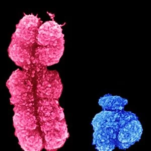
Coloured SEM of egg & sperm during fertilisation
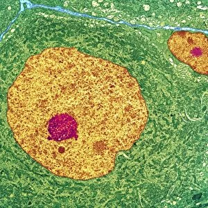
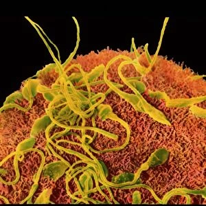
Fertilisation. Coloured Scanning Electron Micro- graph (SEM) of a human egg (ovum) being penetrated by sperm during fertilisation. Sperm are coloured yellow, each with a rounded head and a long tail. They are seen on the thick spongy surface of the zona pellucida which surrounds the egg. The zona pellucida attracts sperm to the egg and enables the sperm to attach. The human female usually produces a single large egg, and only one male sperm can penetrate the eggs wall to fuse with the egg nucleus. When this happens, the egg membrane forms a barrier to any other sperm. Magnification: x960 at 6x7cm size. x1, 260 at 4x5ins
Science Photo Library features Science and Medical images including photos and illustrations
Media ID 6455023
© PROFESSOR P.M. MOTTA ET AL/SCIENCE PHOTO LIBRARY
Egg And Sperm Fertilisation Fertilization Oocyte Ovum Re Production Sperm Zona Pellucida
FEATURES IN THESE COLLECTIONS
> Animals
> Mammals
> Muridae
> Long-tailed Mouse
> Europe
> United Kingdom
> Scotland
> Highlands
> Alness
EDITORS COMMENTS
This print showcases the intricate process of fertilization, depicting a colored scanning electron micrograph (SEM) of an egg and sperm. The image reveals a human egg, known as an ovum, being penetrated by yellow-colored sperm during the crucial moment of fertilization. Each sperm is characterized by its rounded head and long tail, visible on the thick spongy surface of the zona pellucida that envelops the egg. The zona pellucida plays a vital role in attracting and enabling sperm to attach to the egg. In humans, typically only one large egg is produced by females, and it is fascinating to observe how only one male sperm can successfully penetrate its protective wall to fuse with the egg nucleus. Once this fusion occurs, the egg membrane forms a barrier preventing any other sperms from entering. At an impressive magnification level of x960 at 6x7cm size or x1,260 at 4x5ins size, this SEM provides remarkable details about human reproduction anatomy. It offers valuable insights into various aspects such as oocyte development, fertilization processes within our bodies, and highlights key elements like eggs and sperms involved in successful fertilization. This extraordinary photograph from Science Photo Library serves as a testament to scientific advancements in capturing microscopic wonders while shedding light on fundamental concepts related to human fertility and reproductive biology.
MADE IN THE USA
Safe Shipping with 30 Day Money Back Guarantee
FREE PERSONALISATION*
We are proud to offer a range of customisation features including Personalised Captions, Color Filters and Picture Zoom Tools
SECURE PAYMENTS
We happily accept a wide range of payment options so you can pay for the things you need in the way that is most convenient for you
* Options may vary by product and licensing agreement. Zoomed Pictures can be adjusted in the Cart.