Home > Popular Themes > Human Body
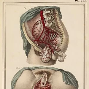
Male groin arteries, 1825 artwork

![]()
Wall Art and Photo Gifts from Science Photo Library
Male groin arteries, 1825 artwork
Male groin arteries. Dissections of a male groin to show areas supplied by the internal iliac artery (red) and its branches. At top, the bladder (left) and rectum (right) hang from the abdominal cavity. At bottom (centred on the anus), the internal pudendal artery supplies the external genitalia. This anatomical artwork is plate 232 from volume 4 of Manuel d anatomie descriptive du corps humain (1825). This 5-volume anatomy atlas was produced by French physician and surgeon Jules Germain Cloquet (1790-1883). The illustrations were by Haincelin. Volume 4 illustrated the anatomy of the circulatory and respiratory systems
Science Photo Library features Science and Medical images including photos and illustrations
Media ID 9222685
© SCIENCE PHOTO LIBRARY
1825 Abdomen Abdominal Abdominal Cavity Anatomical Artwork Anatomical Illustration Anatomy Atlas Arterial System Arteries Axial Bladder Blood Vessels Dissected Dissection French Groin Haincelin Internal Iliac Artery Internal Pudendal Artery Jules Germain Cloquet Lateral Oxygenated Blood Rectum Sagittal Urinary Vascular Volume 4 Volume Iv Anal Artery Blood Vessel Circulatory System Urogenital System
EDITORS COMMENTS
This artwork from 1825 provides a detailed illustration of the male groin arteries, showcasing the intricate network of blood vessels supplied by the internal iliac artery and its branches. Created by French physician and surgeon Jules Germain Cloquet, this anatomical masterpiece is plate 232 from volume 4 of his renowned atlas, Manuel d'anatomie descriptive du corps humain. The image captures a dissected male groin, revealing the areas nourished by the internal iliac artery in vibrant red. At the top, we observe the bladder on the left and rectum on the right hanging from the abdominal cavity. Moving downwards towards the anus, we witness how oxygenated blood is delivered to external genitalia through the internal pudendal artery. This historical artwork not only showcases remarkable artistic skill but also serves as an invaluable resource for understanding human anatomy during that era. It offers insights into our circulatory system's inner workings while shedding light on medical practices prevalent in early 19th-century France. With its meticulous attention to detail and scientific accuracy, this print exemplifies Cloquet's dedication to advancing anatomical knowledge. The collaboration with illustrator Haincelin brings life to each vessel and structure depicted within this composition. As we gaze upon this mesmerizing piece of medical history, it reminds us of our ever-evolving understanding of human physiology and pays homage to those who paved the way for modern medicine.
MADE IN THE USA
Safe Shipping with 30 Day Money Back Guarantee
FREE PERSONALISATION*
We are proud to offer a range of customisation features including Personalised Captions, Color Filters and Picture Zoom Tools
SECURE PAYMENTS
We happily accept a wide range of payment options so you can pay for the things you need in the way that is most convenient for you
* Options may vary by product and licensing agreement. Zoomed Pictures can be adjusted in the Cart.