White matter fibres of the human brain C014 / 5664

![]()
Wall Art and Photo Gifts from Science Photo Library
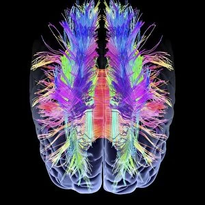
White matter fibres of the human brain C014 / 5664
White matter fibres. Coloured 3D diffusion spectral imaging (DSI) scan of the bundles of white matter nerve fibres in the brain. The fibres transmit nerve signals between brain regions and between the brain and the spinal cord. Diffusion spectrum imaging (DSI) is a variant of magnetic resonance imaging (MRI) in which a magnetic field maps the water contained in neuron fibers, thus mapping their criss-crossing patterns. A similar technique called diffusion tensor imaging (DTI) is also used to explore neural data of white matter fibres in the brain. Both methods allow mapping of their orientations and the connections between brain regions: Data/software: NIH Human Connectome Project /humanconnectomeproject.org)
Science Photo Library features Science and Medical images including photos and illustrations
Media ID 9225605
© PASIEKA/SCIENCE PHOTO LIBRARY
Brain Scan Connections Diffusion Spectral Imaging Diffusion Tensor Imaging Dsi Scan Dti Scan Fibers Fibre Tracking Fibres Human Brain Magnetic Resonance Imaging Mapping Mri Scan Nerve Bundles Nerve Fibre Pathway Pathways White Matter Nervous System
EDITORS COMMENTS
This print showcases the intricate network of white matter fibres in the human brain. Using a cutting-edge technique called diffusion spectral imaging (DSI), this coloured 3D scan reveals the complex pathways through which nerve signals travel between different regions of the brain and even to the spinal cord. By employing magnetic resonance imaging (MRI) technology, DSI maps the water content within these neuron fibers, allowing for a detailed visualization of their criss-crossing patterns. Similar to diffusion tensor imaging (DTI), DSI enables scientists to explore and understand neural data related to white matter fibres in our brains. Both methods provide invaluable insights into mapping their orientations and connections between various brain regions. This particular image is part of an extensive dataset provided by the NIH Human Connectome Project, which aims to unravel the mysteries of our interconnected nervous system. The vibrant colors and intricate details captured in this photograph highlight not only the beauty but also the complexity of our brain's inner workings. It serves as a reminder that beneath its surface lies a vast network responsible for transmitting vital information throughout our bodies. As we continue to delve deeper into understanding these pathways, we unlock new possibilities for diagnosing and treating neurological disorders that affect millions worldwide. Photo credit: PASIEKA/SCIENCE PHOTO LIBRARY
MADE IN THE USA
Safe Shipping with 30 Day Money Back Guarantee
FREE PERSONALISATION*
We are proud to offer a range of customisation features including Personalised Captions, Color Filters and Picture Zoom Tools
SECURE PAYMENTS
We happily accept a wide range of payment options so you can pay for the things you need in the way that is most convenient for you
* Options may vary by product and licensing agreement. Zoomed Pictures can be adjusted in the Cart.