Home > Arts > Artists > G > Thomas Gray
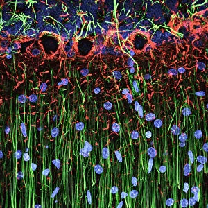
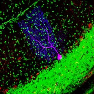
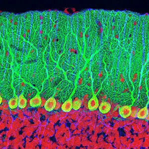
Purkinje nerve cells in the cerebellum

![]()
Wall Art and Photo Gifts from Science Photo Library
Purkinje nerve cells in the cerebellum
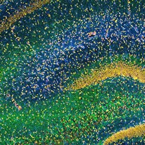
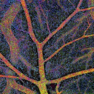
Purkinje cells in the cerebellum. Fluorescent light micrograph of Purkinje cells (green) in the cerebellum of the brain. Purkinje nerve cells have a flask-like body from which numerous highly branched dendrites extend. They are found in the grey matter (cortex) of the cerebellum, at the boundary between the granular layer (blue red) and the molecular layer (red green). The dendrites relay signals to the cell body, which passes them on through its single axon (green) in the granular layer. The cerebellum is a structure at the base of the brain that plays an important role in motor control, sensory perception and learning. Magnification: x380 when printed 10cm wide
Science Photo Library features Science and Medical images including photos and illustrations
Media ID 6448951
© THOMAS DEERINCK, NCMIR/SCIENCE PHOTO LIBRARY
Cerebellar Cerebellum Cortex Dendrite Dendrites Fluorescent Light Micrograph Granular Layer Gray Grey Matter Histological Histology Molecular Layer Nerve Cell Neuron Purkinje Cell Brain Light Microscope Nervous System Neurological Neurology
FEATURES IN THESE COLLECTIONS
> Animals
> Mammals
> Muridae
> Blue-grey Mouse
> Arts
> Artists
> G
> Thomas Gray
> Science Photo Library
> Specialist Imaging
EDITORS COMMENTS
This print showcases the intricate beauty of Purkinje nerve cells in the cerebellum. The fluorescent light micrograph reveals these remarkable cells, with their distinct flask-like bodies and highly branched dendrites extending outwards. Positioned at the boundary between the granular layer and molecular layer in the grey matter of the cerebellum, these green-hued Purkinje cells play a crucial role in relaying signals. The image provides a glimpse into the complex inner workings of our brain's structure. With its single axon visible amidst the granular layer, this photograph highlights how these nerve cells transmit information from their dendrites to their cell body before passing it on further. The cerebellum itself holds immense significance as a vital component responsible for motor control, sensory perception, and learning. This microscopic view emphasizes its importance within our nervous system. With a magnification of x380 when printed 10cm wide, this print offers an up-close exploration into neurology and histology. It serves as a reminder of both our biological intricacies and the wonders that lie within us all.
MADE IN THE USA
Safe Shipping with 30 Day Money Back Guarantee
FREE PERSONALISATION*
We are proud to offer a range of customisation features including Personalised Captions, Color Filters and Picture Zoom Tools
SECURE PAYMENTS
We happily accept a wide range of payment options so you can pay for the things you need in the way that is most convenient for you
* Options may vary by product and licensing agreement. Zoomed Pictures can be adjusted in the Cart.