Trachea lining, TEM C014 / 1472

![]()
Wall Art and Photo Gifts from Science Photo Library
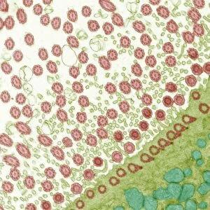
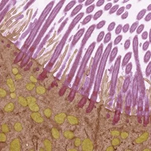
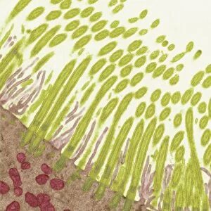
Trachea lining, TEM C014 / 1472
Trachea lining. Transmission electron micrograph (TEM) of a transverse section through the lining of the trachea (windpipe), which links the larynx (voicebox) to the lungs. The epithelial lining consists of cells that have hair-like cilia (yellow, upper left) on their surface and mucus-secreting glands. Mucus secreted by the glands traps debris, such as dust particles or bacteria, in the inhaled air, while the beating of the cilia moves the mucus and particles upwards out of the respiratory tract, helping to keep the lungs and airways clear and prevent infection. Clear basal bodies (yellow rings) and mitochondria (purple) are visible in the cell bodies. Magnification: x7000 when printed 10 centimetres wide
Science Photo Library features Science and Medical images including photos and illustrations
Media ID 9226399
© STEVE GSCHMEISSNER/SCIENCE PHOTO LIBRARY
Bodies Cilia Cilium Cytological Cytology Gland Glands Histological Histology Lining Microvilli Microvillus Mitochondria Mitochondrion Mucous Cell Mucus Respiratory System Secretory Trachea Transmission Electron Micrograph Transmission Electron Microscope Transverse Section Windpipe Sectioned
EDITORS COMMENTS
This print showcases the intricate lining of the trachea, captured through a transmission electron microscope. The trachea, also known as the windpipe, serves as a vital pathway connecting the larynx to the lungs. In this transverse section image, we can observe the remarkable epithelial lining composed of specialized cells adorned with hair-like cilia and mucus-secreting glands. The yellow-colored cilia located in the upper left corner play a crucial role in maintaining respiratory health. As air is inhaled, mucus secreted by these glands acts as a sticky trap for harmful debris like dust particles or bacteria present in our environment. Simultaneously, the beating motion of these cilia propels both mucus and trapped particles upwards and out of our respiratory tract. Notably visible within each cell are clear basal bodies resembling yellow rings alongside purple mitochondria responsible for energy production. These cellular components contribute to sustaining proper functioning within this dynamic system. With an impressive magnification level of x7000 when printed at 10 centimeters wide, this photograph allows us to marvel at nature's intricacy on a microscopic scale. It serves as a testament to how our body employs ingenious mechanisms to keep our lungs and airways clear while safeguarding against potential infections. Photographer Steve Gschmeissner from Science Photo Library has masterfully captured this snapshot that encapsulates both beauty and scientific significance found within human biology – reminding us once again of nature's awe-inspiring complexity.
MADE IN THE USA
Safe Shipping with 30 Day Money Back Guarantee
FREE PERSONALISATION*
We are proud to offer a range of customisation features including Personalised Captions, Color Filters and Picture Zoom Tools
SECURE PAYMENTS
We happily accept a wide range of payment options so you can pay for the things you need in the way that is most convenient for you
* Options may vary by product and licensing agreement. Zoomed Pictures can be adjusted in the Cart.