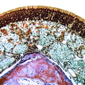
Nose mucosa, light micrograph

![]()
Wall Art and Photo Gifts from Science Photo Library
Nose mucosa, light micrograph
Nose mucosa. Light micrograph of a section through the nasal mucosa, the tissue that lines the airways of the nose. Air flows through hair-like cilia, found on the upper surface of epithelial cells (pink, top), which form the top layer of the nasal mucosa. The cilia are coated in sticky mucus and serum produced by the Bowmans glands (red) in the underlying mucous membrane (yellow). These secretions serve to trap dust and pathogens. Beneath the mucous membrane is a layer of supportive cartilage (orange) and then bone tissue (purple)
Science Photo Library features Science and Medical images including photos and illustrations
Media ID 6449147
© STEVE GSCHMEISSNER/SCIENCE PHOTO LIBRARY
Cartilage Ciliated Epithelial Epithelium False Colour Glands Goblet Histological Histology Mucosa Mucous Mucous Membrane Mucus Nasal Nasal Cavity Nose Olfaction Olfactory Secretion Sense Serum Smell Supportive Tissue Tissue Bowman Cells False Coloured Light Micrograph Light Microscope
MADE IN THE USA
Safe Shipping with 30 Day Money Back Guarantee
FREE PERSONALISATION*
We are proud to offer a range of customisation features including Personalised Captions, Color Filters and Picture Zoom Tools
SECURE PAYMENTS
We happily accept a wide range of payment options so you can pay for the things you need in the way that is most convenient for you
* Options may vary by product and licensing agreement. Zoomed Pictures can be adjusted in the Cart.